Inside the clinic
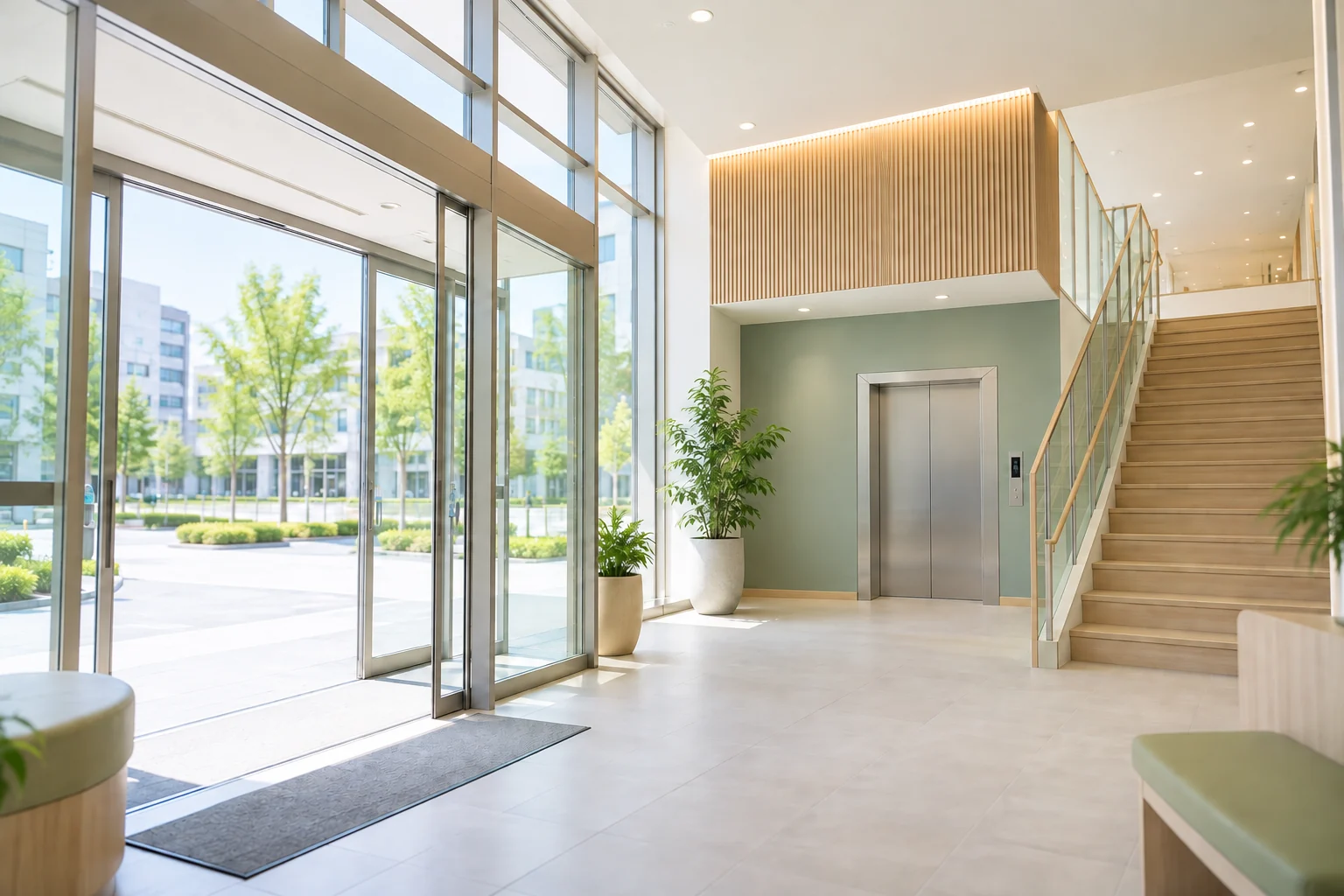
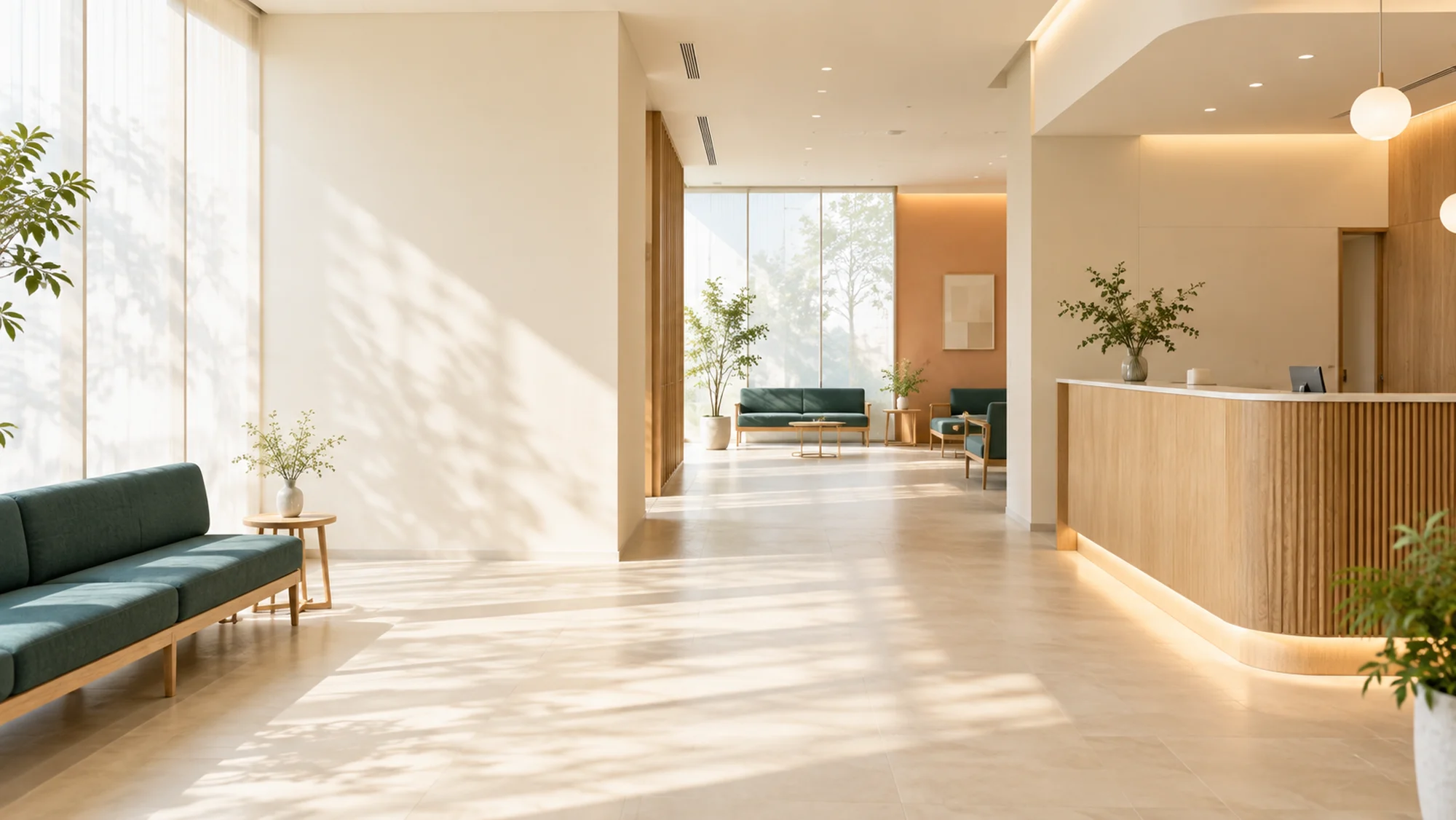
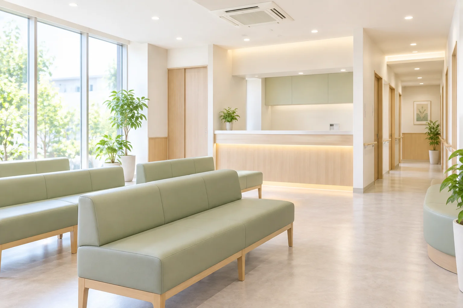
We are on the second floor of Hiwadai Medical Mall, just outside the elevator. Reception and the waiting area are separated from the examination-room route, so patients moving between tests do not need to pass through the waiting area.
There are 22 waiting-room seats, including four higher seats with armrests for people who find standing difficult. Facilities include a wheelchair-accessible toilet with handrails, a baby seat and a children's area. There are no steps from the entrance to the consultation room.
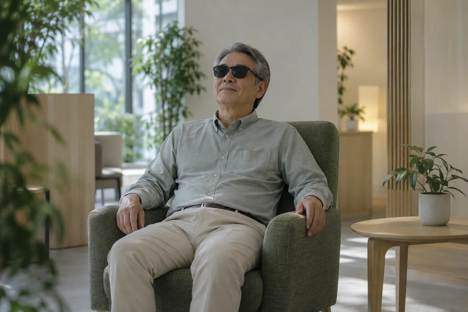
During the 2024 renovation, we added a dimly lit rest area for people experiencing glare after examinations. You can rest there after pupil dilation until your vision settles.
Our team
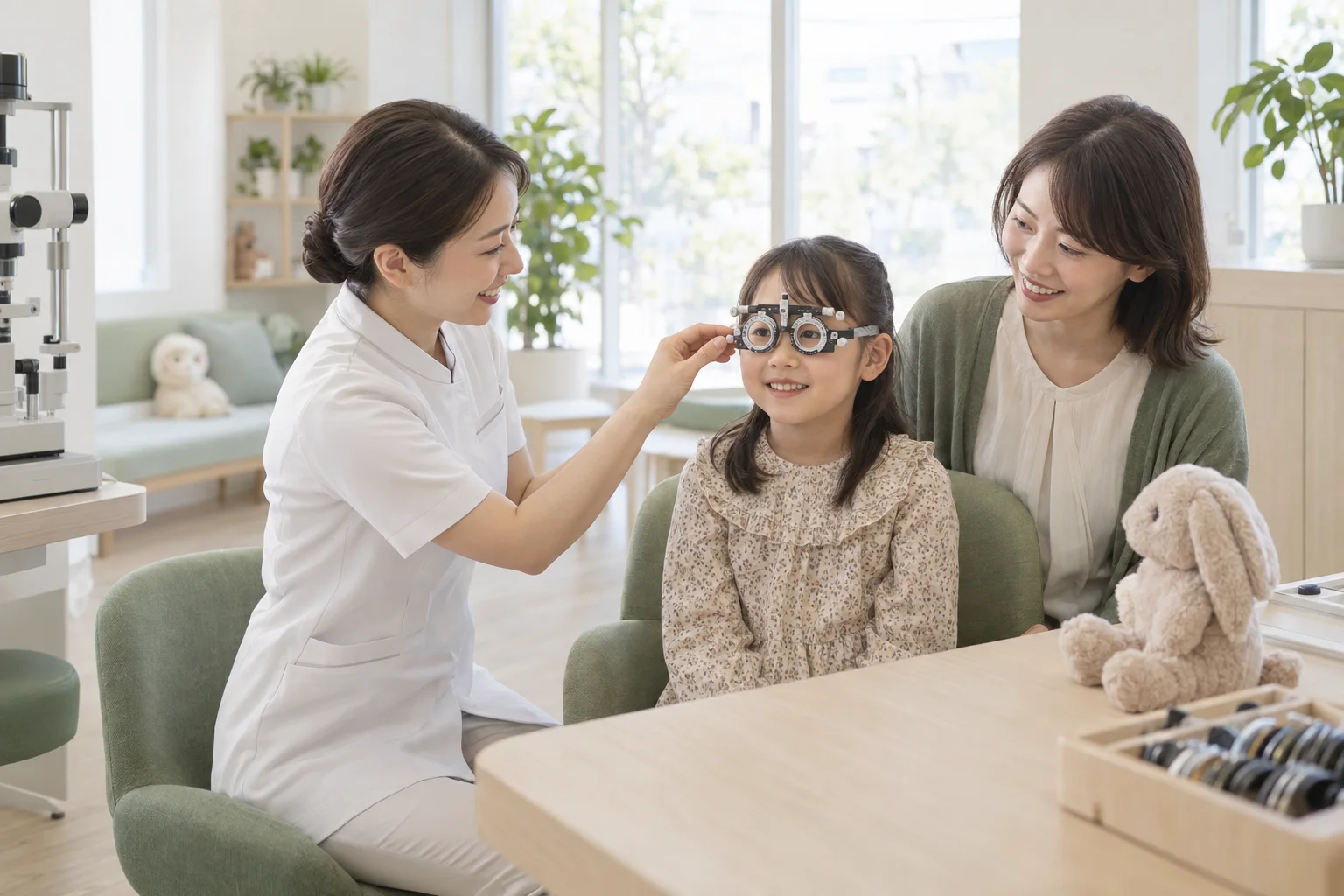
Our team includes two full-time orthoptists (specialists in vision testing and eye coordination), two nurses, one part-time orthoptist and three reception and medical administration staff.
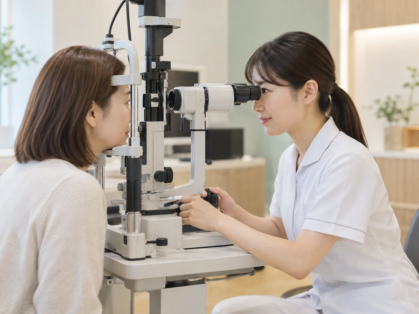
Orthoptists carry out visual acuity, visual field and eye alignment tests. We have a dedicated practitioner for children's amblyopia training and strabismus testing, where experience affects results. On surgery days, two nurses and one orthoptist work in the operating room.
Optical coherence tomography (OCT)
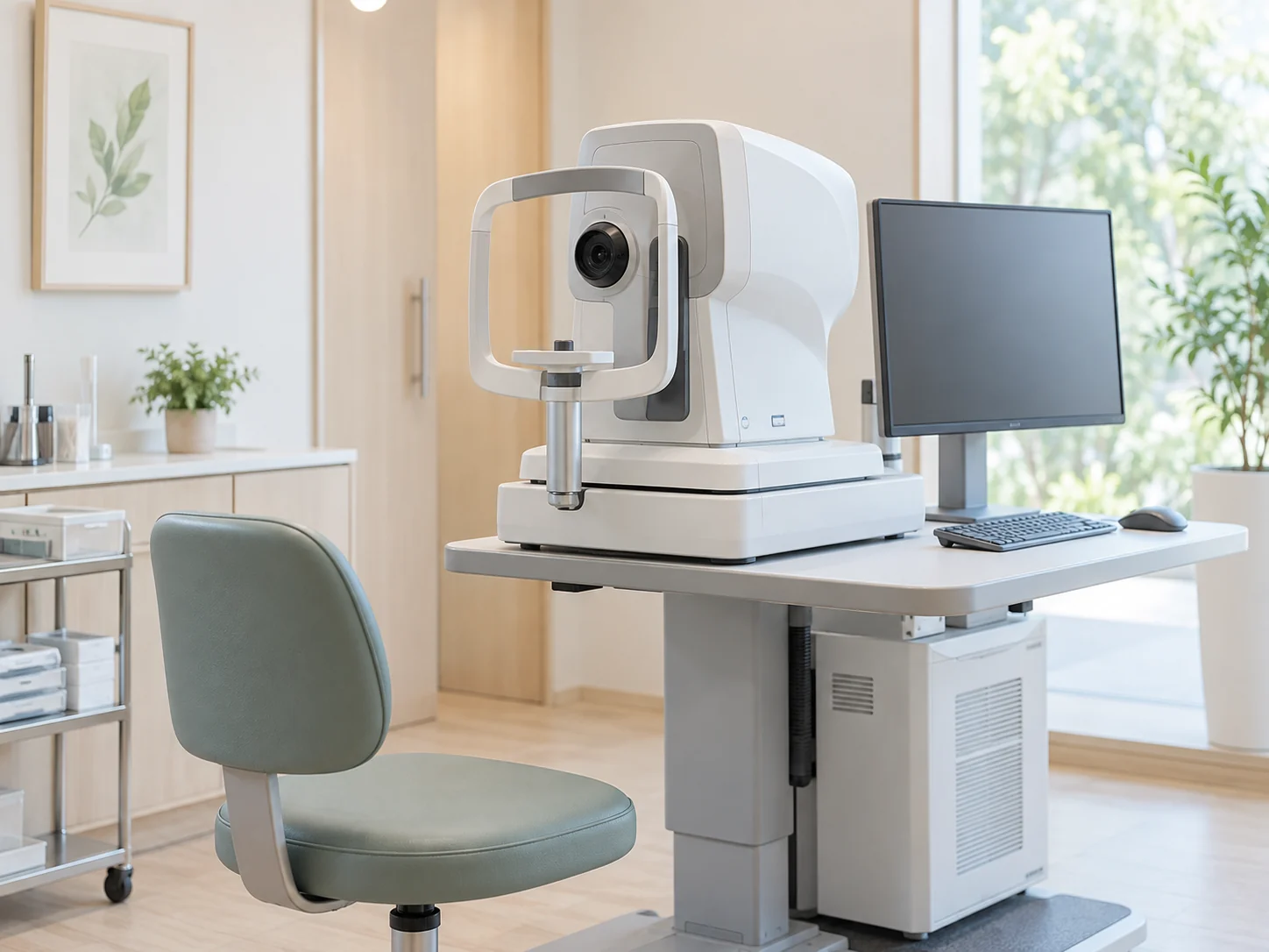
OCT uses infrared light and analyzes its reflection to create cross-sectional images of the retina. It shows the retina's roughly ten-layer structure in detail.
For glaucoma, we measure nerve fiber thickness and compare changes with earlier scans. For age-related macular degeneration or diabetic macular edema, we check the location and thickness of swelling or bleeding. A scan takes about 10 seconds per eye and does not touch the eye.
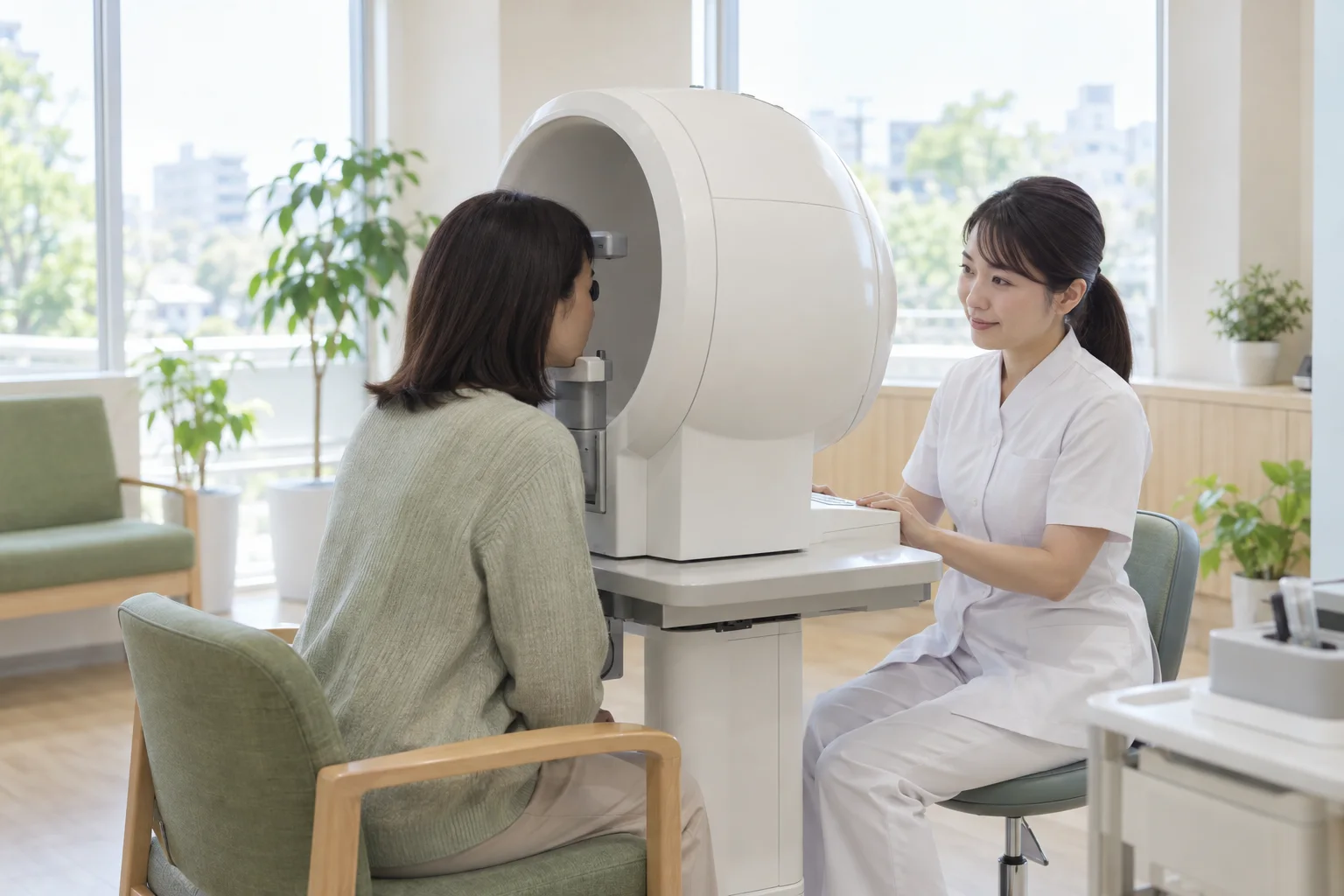
Automated visual field testing
This test checks where you can see points of light, measuring your field of vision and the retina's sensitivity to light. It helps diagnose glaucoma and assess how well treatment is limiting progression.
Glaucoma-related field loss often starts away from the center and may go unnoticed when both eyes are open. For many patients, this test first reveals the loss. It takes about 10-15 minutes per eye.
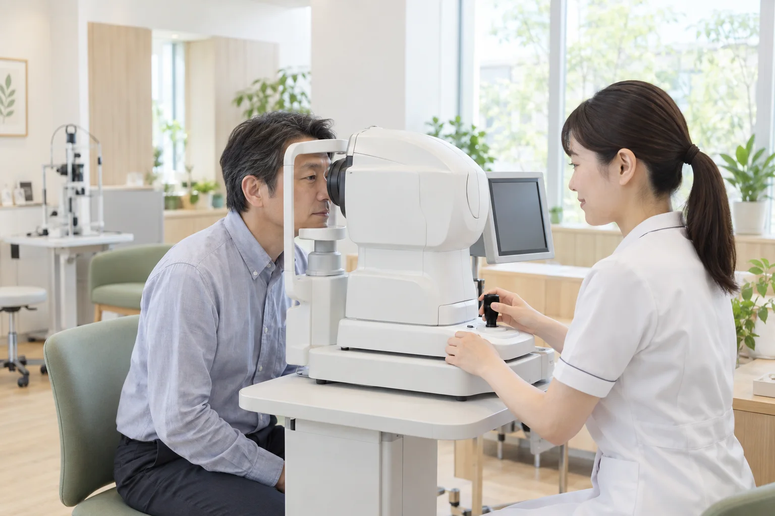
Non-mydriatic retinal camera
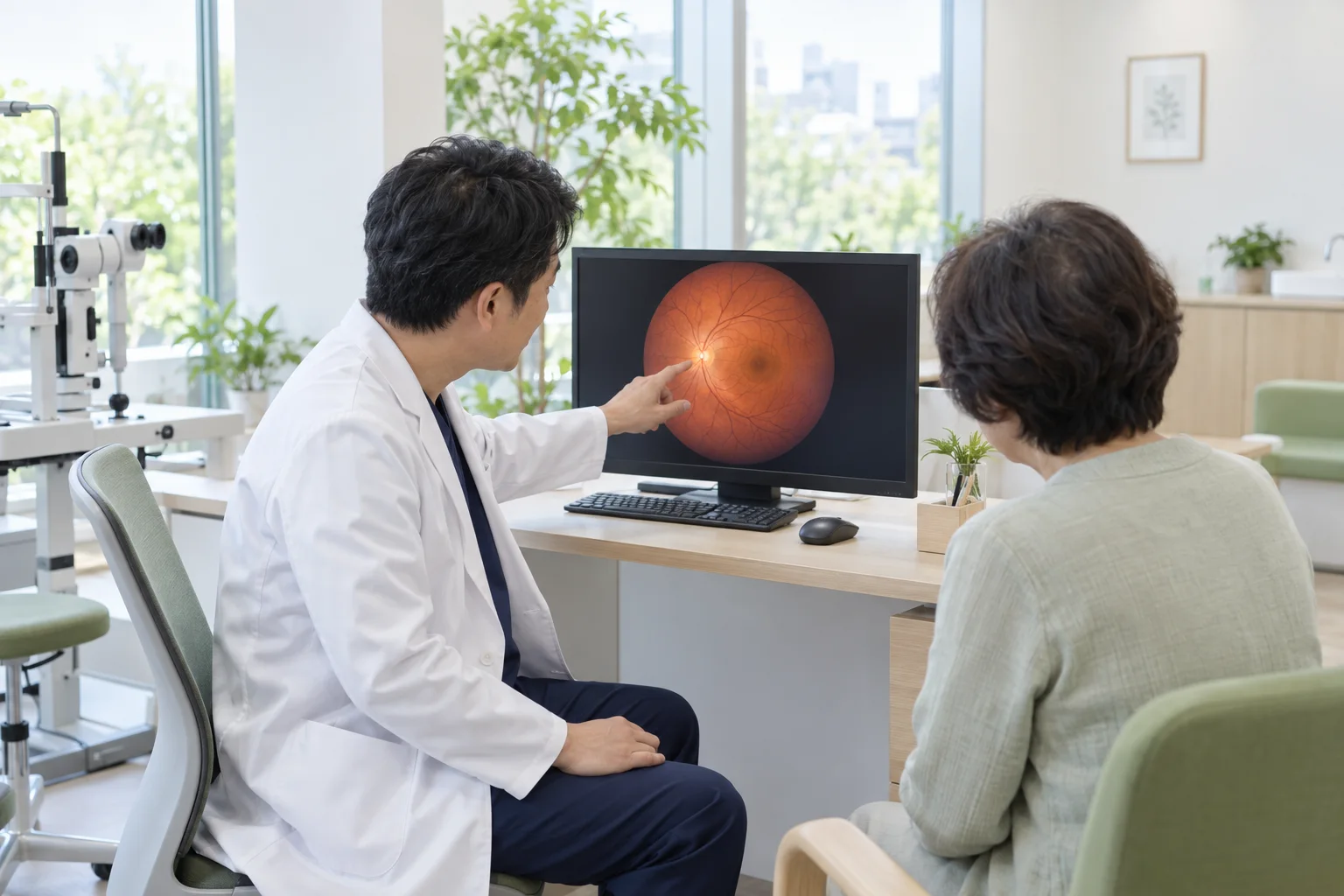
This camera photographs the back of the eye without pupil-dilating drops. It records retinal blood vessels, the shape of the optic disc, and any bleeding or white spots.
Images are taken under consistent conditions and saved so we can compare changes over several years. When a detailed view of the peripheral retina is needed, we use a dilated retinal examination instead.
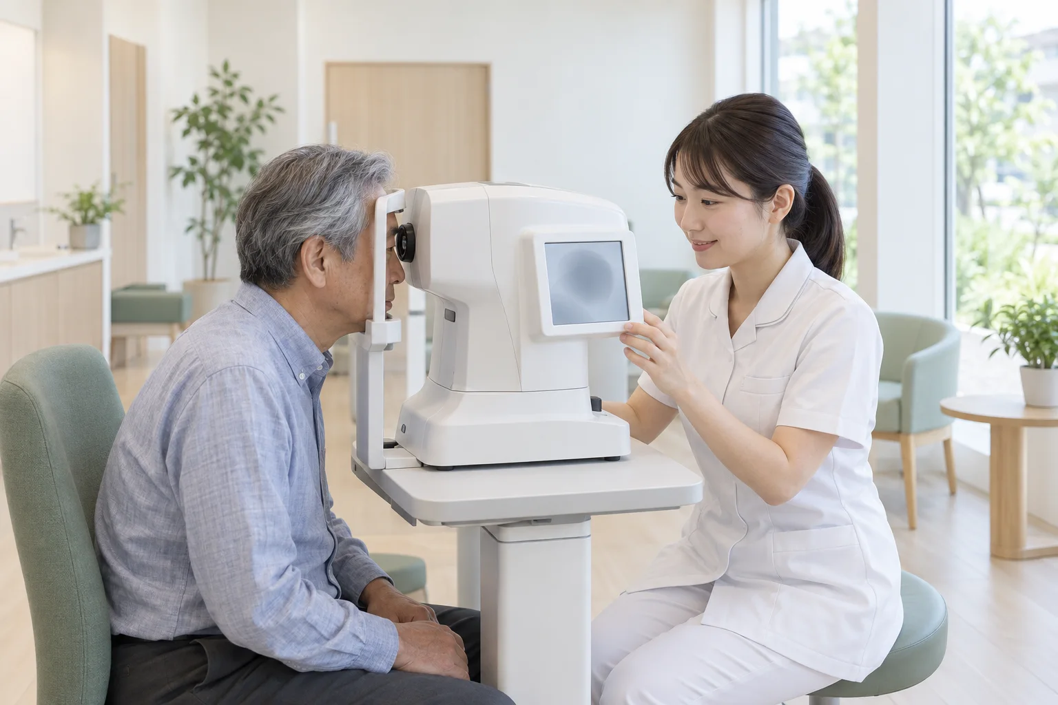
Specular microscopy
This examination records the number and shape of endothelial cells on the inner surface of the cornea. These cells keep the cornea clear but do not regenerate once lost.
If the number falls below a certain level, the cornea may swell and become cloudy. We check these cells before cataract surgery and during regular examinations for long-term contact lens wearers.
Other examination equipment
- Non-contact tonometer
- Measures eye pressure with a puff of air, for glaucoma care
- Autorefractor and keratometer
- Measures short-sightedness, long-sightedness, astigmatism and corneal curvature
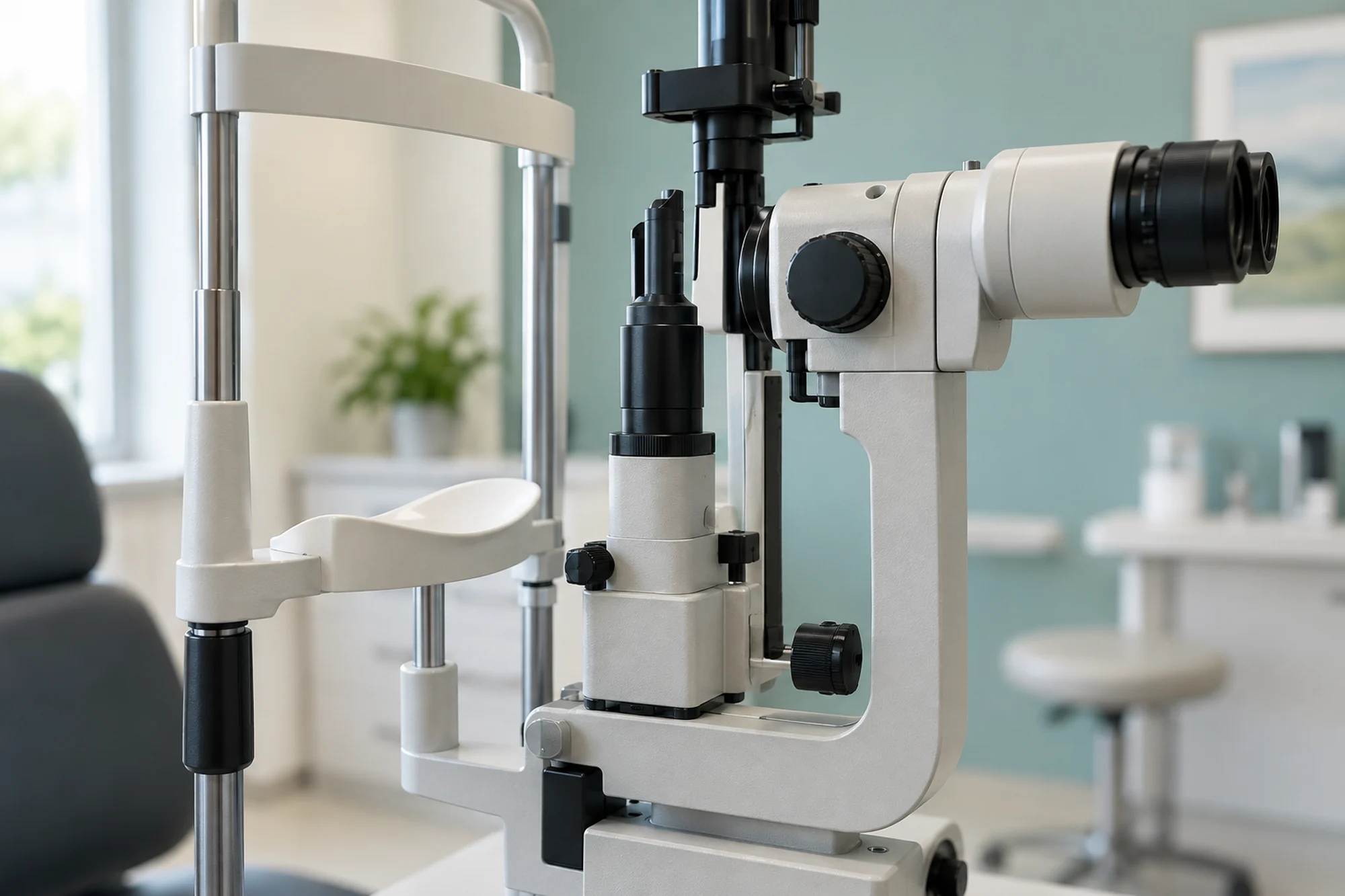
- Slit-lamp microscope
- Magnifies the cornea, conjunctiva and lens
- Optical biometer
- Determines the power of the intraocular lens for cataract surgery
- Laser photocoagulator
- Treats diabetic retinopathy and retinal tears
- YAG laser
- Treats clouding of the posterior capsule after cataract surgery